Boparai, Nikpreet: A Structural Comparison of Wild-Type and S42Y Mutant Alpha-Synuclein Fibrils
Title: A Structural Comparison of Wild-Type and S42Y Mutant Alpha-Synuclein Fibrils
Name: Nikpreet Boparai
Major: Cell Biology and Neuroscience
School affiliation: School of Arts and Sciences
Programs: Division of Life Sciences Summer Undergraduate Research Fellowship (DLS-SURF)
Other contributors: Jennifer Jiang, Muyuan Chen, Zhiyong Bai, Yoon-Seong Kim, Wei Dai
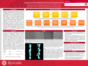
Abstract: Aggregation of alpha-synuclein (⍺-syn) plays a crucial role in the pathophysiological progression of Parkinson’s disease, a neurodegenerative disorder that involves degeneration of dopaminergic neurons in the substantia nigra of the basal ganglia, leading to both motor and non-motor deficits. Alpha-synuclein is a small, soluble, intrinsically disordered protein (IDP) that is encoded by the SNCA gene and has various physiological functions including microtubule-associated activity, neuronal plasticity, antioxidant activity in membranes, and SNARE complex assembly. It is predominantly expressed in neural tissue and is the main component of Lewy bodies and neurites, which are histopathological signatures of Parkinson’s disease and related synucleinopathies such as dementia with Lewy bodies (DLB) and multiple system atrophy (MSA). There is a connection between the aggregation of ⍺-syn and the onset and progression of Parkinson’s disease, yet the molecular mechanisms as to how these aggregates lead to neurodegeneration remain elusive. This project is focused on investigating the structural basis underlying ⍺-syn aggregation and fibrillation propensity. Alpha-synuclein is exceedingly complex and forms heterogenous conformational states which contribute to the multi-faceted nature of Parkinson’s disease. Certain mutations and environmental factors influence the conformation of the protein which in turn influences the kinetics of aggregation. The goal of this project is to perform a structural analysis of ⍺-syn preformed fibrils (PFFs) from the wild type and a post-translational mutant S42Y. This serine to tyrosine substitution results in enhanced aggregation properties. We will use cryo-electron tomography (cryoET) and computational subtomogram analysis to resolve 3D ultrastructures of WT and S42Y PFFs. By comparing 3D structures of WT PFFs and S42Y PFFs, we aim to elucidate the structural basis underlying the variation of aggregation kinetics in the S42Y mutant.