Lei, Shunyao: Isolation and Characterization of Exosomes from Human Amniotic Membranes

Title: Isolation and Characterization of Exosomes from Human Amniotic Membranes
Name: Shunyao Lei
Major: Biomedical Engineering
School affiliation: School of Engineering
Programs: Aresty – Research or Conference Funding Recipient
Other contributors:
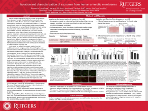
Abstract: Human amniotic membrane (AM) is an inner surrounding of the embryo and is made of epithelial cells, stromal cells and extracellular matrix. Recent studies showed that AM has anti-inflammatory, anti-fibrotic, and antimicrobial effects. In a previous research done in our lab, the preserved viable amnion showed anti-fibrotic activity on human fibrotic fibroblasts and such activity is mostly due to factors released from the viable cells in the preserved AM. However, the identity of such factors and the mechanism of action of anti-fibrotic activity remained to be explored. Exosome is nanoscale vesicle produced in most cells and gets involved in the communications between cells through the transfer of DNA, RNA, and proteins. Studies showed that exosomes are vital in the delivery of necessary components for fibrosis regression and help with the inactivation of hepatic stellate cells. We hypothesized that exosomes from AM may be the mediators for the anti-fibrotic activity of AM. In this study, we isolated nano-sized vesicles from AM conditioned medium using sequential ultracentrifugation and filtration methods. These vesicles were characterized using particle size analysis (Dynamic Light scattering), transmission electron microscopy and Western blotting. These vesicles showed the characteristics of exosomes with spherical morphologies in size range of 70-90 nm and enriched with tetraspanins such as (CD9, CD63 and CD81). Furthermore, the anti-fibrotic activity of AM-derived exosomes was evaluated in human hepatic stellate cells (LX-2), which is an in vitro model for studying fibrosis. The proliferation of LX-2 cells in the presence or absence of the exosomes was monitored using alamarBlue assay and the migration of cells was measured using a scratch wound assay. While AM-derived exosomes had no effect on the proliferation of the resting LX-2 cells, these exosomes inhibited the proliferation of TGFB1-activated LX-2 cells. The presence of exosomes also reduced the migration of the LX-2 cells. These results suggested that exosomes are released from AM tissue and may carry out the anti-fibrotic activity from the tissue to their target LX-2 cells.