What We Do
In order to establish a full picture of out veterans we incorporate a variety of tests during evaluations. While we have multiple types of tests to choose from, and we are learning and adding to our tool box constantly.

-
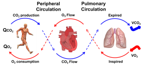
A cardiopulmonary exercise test (CPET) is an evaluation the pulmonary, cardiovascular and skeletal muscle systems.
More specifically, this tests looks at how well these three systems work together and how they respond to a maximal exercise test. Their ability to respond adequately to this stress is a measure of their physiologic competence.
This test can be performed on a treadmill or on a stationary bike.
-
Pulmonary function tests (PFTs) are tests that show how well the lungs are working. By performing multiple maneuvers, all of which are meant to stress the lungs in a particular way, this tests allows us to measure lung volume, capacity, rates of flow, and gas exchange.
-
This test is performed to further measure lung function. This tests sends sound waves down the lungs. Based on how the waves travels in the lungs, a mathematical analyses of resistance and reactance of the lungs.
FOTs are an easy to examine deep airways in your lungs
-
This tests allows us to measure how well the lungs can transfer oxygen to circulation blood cells. By giving the subject a known concentration of tracer gases, that bind to blood cells better than oxygen, we can measure the concentrations inhaled and exhaled to evaluate diffusion capacity. Our lab uses two different tracer gases for our tests.
- Carbon Monoxide (DLCO)
- Nitric Oxide (DLNO)
-
A stress echocardiography, is another test used to evaluate the pulmonary, cardiovascular and skeletal muscle systems.
In this test images of the heart and blood vessels around it are obtained using an ultrasound.
In our lab this test is performed on a tilt bike which allows the sonographer to acquire live images of the heart while exercising.
-
Flow mediated dilation tests look at how well your arteries react to increase blood flow. This test is useful in evaluating the endothelial dysfunctions. In our lab we evaluate changes in the brachial artery using an ultrasound and inducing dilation in two different ways.
- Cuff: We restrict blood flow using an arm cuff. Endothelial functions are evaluated by measuring changes in the artery after pressure release.
- Nitroglycerin: We administer the vasodilatory (nitroglycerin) and measure changes in the artery in real time.
-
Compared to more traditional CT scans HRCTs can obtain extremely thin slices and multiple images to help establish a better profile of the lung.
HRCT routinely uses these very thin slices (0.5mm – 2mm) to effectively clarify obstructive or restrictive patterns in the small airways and lung parenchyma.
-
The Seahorse XFp analyzer is used to measure the metabolic ability of cells.
By measuring O2 consumption and proton release the XFp allows a real-time picture of cellular respiration.
We can learn about the quality of the mitochondria population isolated by looking at the extracellular flux.