Youssef, Mark: Alzheimer’s Disease Effect on Human Neuron Axon Initial Segment
Title: Alzheimer’s Disease Effect on Human Neuron Axon Initial Segment
Name: Mark Youssef
Major: Biological Sciences
School affiliation: School of Environmental and Biological Sciences
Programs: Aresty Summer Science Program
Other contributors: Dina Popova, Petronio Zalamea, Ronald Hart
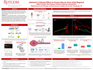
Abstract: Axonal death has been reported in Alzheimer’s Disease (AD) mouse models, yet there has been very little data on AD human neurons. One of the major hypotheses in AD research is that the accumulation of an insoluble neurotoxin, amyloid β-protein (Aβ), in the brain is the main influencer for AD. The Axon Initial Segment (AIS), is a compartment near the base of the axon, which generates and shapes the action potential using the voltage-gated potassium channels (Kv) and voltage-gated sodium channels (Nv) which are concentrated at the AIS by anchoring to the microtubules using a protein called Ankyrin G (Pan et al., 2006; Cooper, 2011; Xu and Cooper, 2015; Meza et al., 2017). Previous studies suggested that the Ankyrin G levels are downregulated in AD transgenic mice models (Sun et al., 2014). Therefore, we hypothesized that human neurons treated with oligomeric Aβ would cause a downregulation in Ankyrin G which would cause the AIS to be smaller, farther from the soma, and alter neuronal responsiveness. One of the models used in AD research is the Swedish mutation of APP (APPswe), which has been known to increase cleavage of the amyloid precursor protein (APP) to increase secretion of Aβ. We have constructed a lentiviral vector expressing human APPSwe for the secretion of oligomeric Aβ by Human Embryonic Kidney (HEK) cells. Once we have mature human induced pluripotent stem cell (iPSC) neurons, we use conditioned medium from the HEK cells to treat the neurons for 2 days. This is followed by immunofluorescence staining to tag the AIS and the neuron processes using the Ankyrin G and the Beta-III Tubulin antibodies. After the neurons are stained, imaging is done by confocal microscope. These images are then analyzed using image analysis software (FIJI) for axonal morphology and the distance from SOMA. The results show significant differences in the AIS between the APPswe treatment and Wild Type. These results are significant as this is the first observed effect of Aβ on human neurons. This information suggests that AD utilizes Aβ to decrease neuronal survivability.