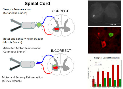
Gradients to guide axon growth
During development, a myriad of chemical and mechanical signals guide axon growth to the specific motor or sensory target. In our lab, we aim to recapitulate these signals in vitro to identify combinations of bioactive and mechanical signals that increase neurite outgrowth, and/or bias directionality of outgrowth. Control of neurite outgrowth and direction of growth using identified biochemical and/or mechanical patterns may translate to an increased potential for a successful biomaterial containing these signals. This biomaterial can be utilized for conditions such as peripheral nerve and spinal cord injuries, where it is imperative that axons accurately reconnect to their previous targets.

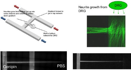
We developed an in vitro microfluidic system with two inlets which we can inject combinations of modified or unmodified type-I collagen hydrogels. These hydrogels may contain gradients of covalently grafted bioactive peptides, including laminin fragments such as IKVAV and YIGSR, or of mechanical stiffness using a natural crosslinker such as genipin. In vitro results using dorsal root ganglia explants from E8 chick embryos have demonstrated that neurite outgrowth is biased down a gradient of stiffness compared to its converse. Neurites also grow significantly longer up steep gradients of YIGSR, shallow gradients of IKVAV, and in combination compared to unmodified collagen controls.

Using these cost-effective, simple in vitro microfluidic techniques, we have identified combinations of bioactive and mechanical elements that are advantageous to test in vivo to control neurite outgrowth.

Photo-active, Thermoreversible Type-I Collagen

Type-I collagen hydrogels are often used for soft tissue engineering applications as they self-assemble into a fibrillar gel in vivo and are biocompatible, biodegradable, and naturally support cell adhesion and growth. While these hydrogels are easily amenable to chemical modifications, they have been criticized for their weak mechanical properties and lack of spatial control of bioactive/mechanical patterns. Our lab covalently grafted methacrylic acid to the free amines on the lysine residues of type-I collagen to synthesize collagen methacrylamide (CMA). CMA can be photocrosslinked in the presence of long-wave UV light and a photoinitiator for specific spatiotemporal control of chemical and mechanical patterns.

Additionally, CMA can thermo-reversibly self-assemble, forming a liquid suspension at cool temperatures, and re-assembling into a hydrogel at physiological temperature. Currently, we are using CMA for tissue engineering strategies, such as the design of specific 3-D cell microenvironments and methods for free form fabrication for controlled tissue architectures. For example, below are pictured two photo-masks next to opaque CMA scaffolds generated by exposing CMA gels to UV light through the masks, and then “cold-melting” the unreacted CMA away.

Biomaterials that Induce Preferential Motor Reinnervation
Peripheral nerve injury (PNI) is a common outcome following physical trauma such as motor vehicle accidents, resulting in debilitating loss of motor and sensory function. Direct reconnection of nerve stumps through suturing is preferred in small gaps; however the current gold standard for large gap nerve repair, an autologous nerve graft, is limited by the availability of nerve tissue and donor site morbidity. Nerve guidance conduits present a promising tissue engineered solution, however, currently available commercial nerve guidance conduits are ineffective in regenerating nerve defects larger than 3cm, due in large part to their hollow lumens which serve only to confine axonal growth. Polysialic acid (PSA) and a factor first found on natural killer cells (HNK-1) are two naturally occurring glycans which are naturally upregulated following nerve injury.
PSA enhances neurite outgrowth and Schwaan cell migration, whereas HNK-1 preferentially enhances growth of motor neurons. Research in our lab and with our collaborators has identified small glycomimetic peptides which mimic the activity of PSA and HNK-1. Together these glycomimetic peptides enhance the speed and the quality of regeneration by preferentially targeting the regeneration of motor neurons, a phenomenon known as preferential motor reinnervation. Our lab has demonstrated increased efficacy of these compounds by covalently grafting them to collagen hydrogels. Peptide grafted hydrogels enhanced neurite outgrowth in sensory and motor neurons as well as increased proliferation and extension of Schwaan cells in vitro. Further, these peptide grafted hydrogels promoted functional motor recovery in vivo. Our ongoing efforts are to optimize the presentation of these glycomimetic peptide cues along with structural guidance cues to design a functional conduit filling which serve to accelerate regeneration across a nerve gap.