Image Galery
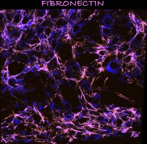
Fibronectin matrix: Sudipto Munshi
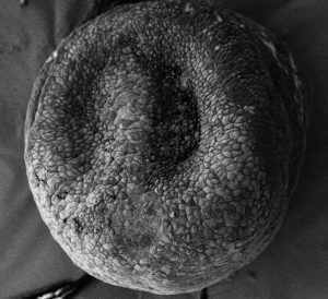
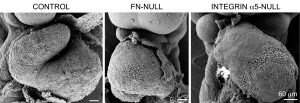
Scanning EM of an early mouse embryo: Sophie Astrof
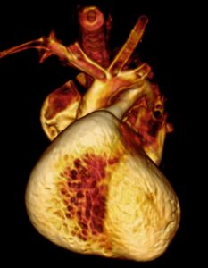
Heart and the aortic arch arteries, imaged by micro computed tomography: AnnJosette Ramirez
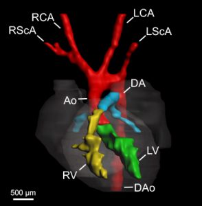
E10.5 embryo heart and vasculature:AnnJosette Ramirez
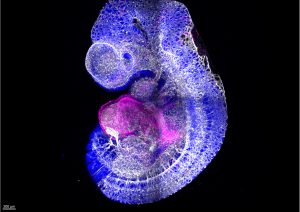
E8.0 mouse embryo: Michael Warkala
Pharyngeal arch arteries: AnnJosette Ramirez
Delicate arch

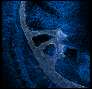
Thick fibronectin fibril at 23 nm resolution
Scanning EM of hearts: Sophie Astrof
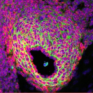
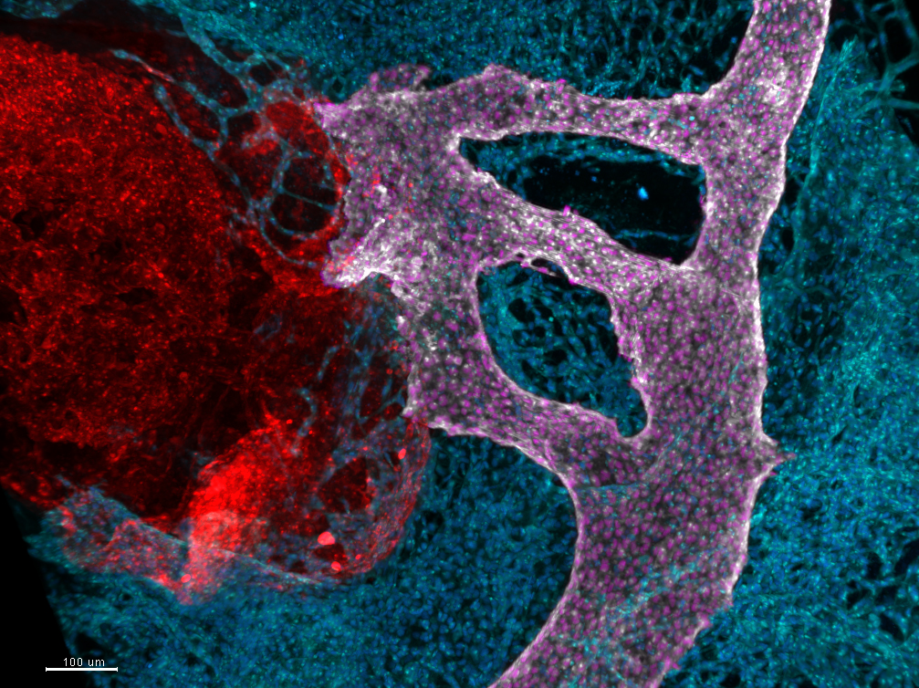
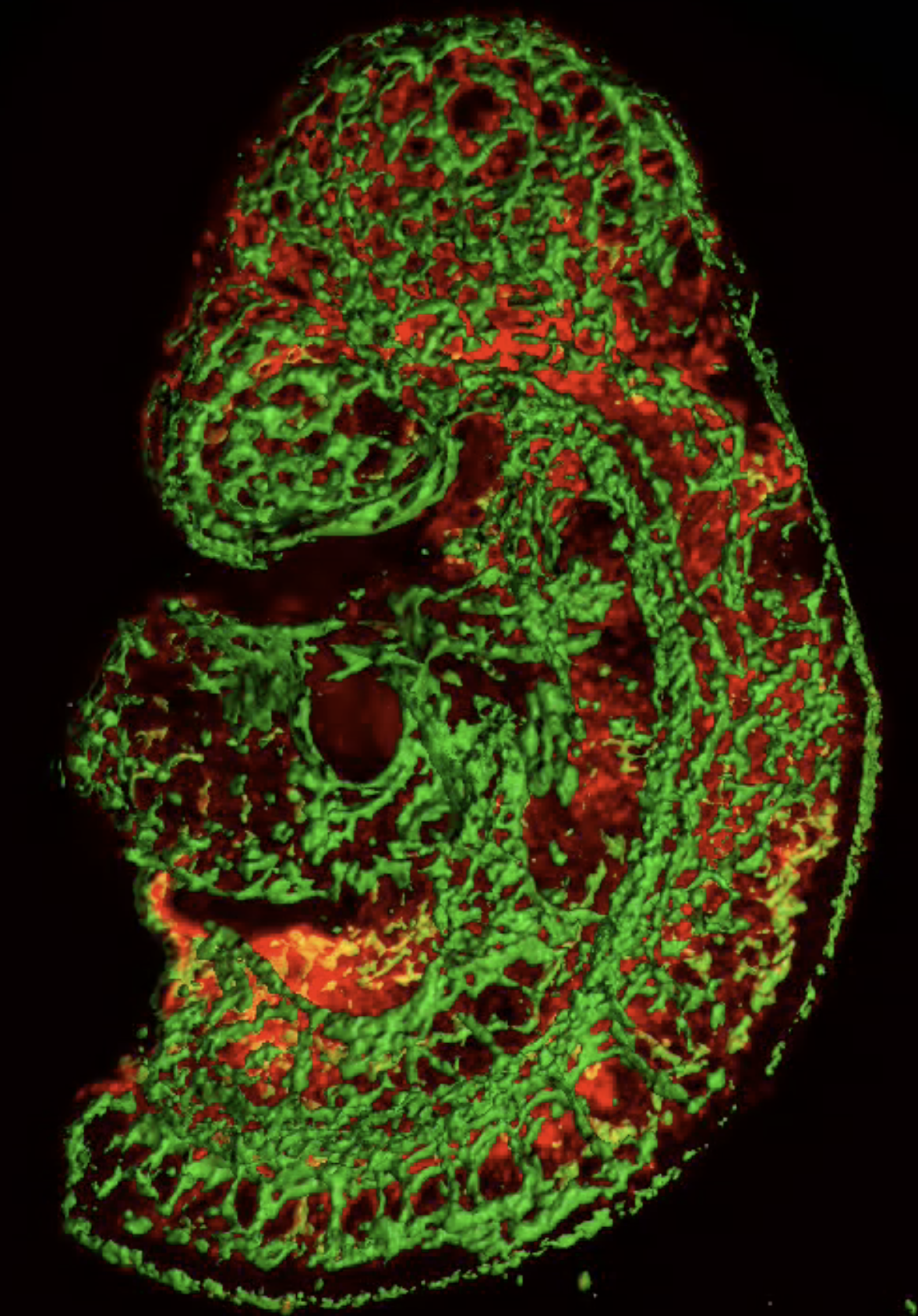
Pharyngeal arch arteries in E10.5 mouse embryo. Sagittal view of three-dimensional maximum intensity projection of whole-mount immunofluorescence staining shows the heart connecting with the dorsal aorta through three pharyngeal arch arteries. Endothelial cell nuclei in the pharyngeal arch arteries and the dorsal aorta are marked in pink by highlighting the expression of ERG. Endothelial cell membranes are marked by the expression of Vascular Endothelial Growth Factor 2 (VEGFR2): red (heart), white (pharyngeal arch arteries and dorsal aorta), and blue (the rest of the embryo). Photo Credit: Michael Warkala
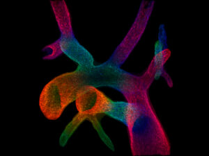
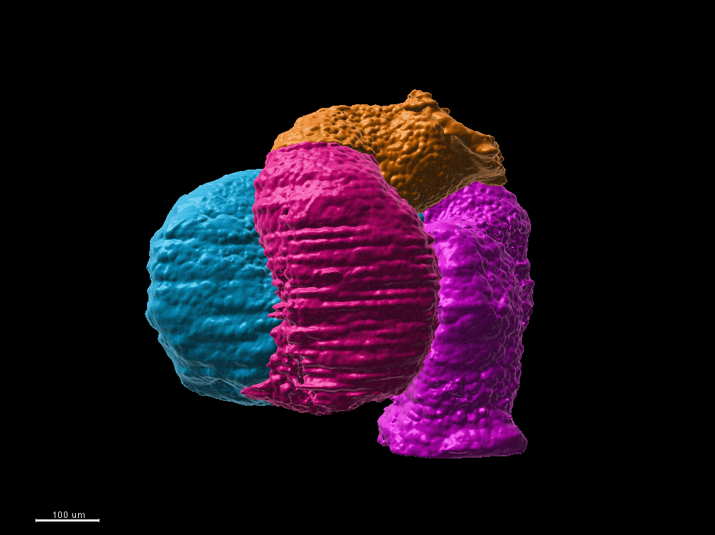
The heart from 9.5-day-old mouse embryo was surfaced to highlight the distal outflow tract (orange), proximal outflow tract and the right ventricle (maroon), the left ventricle and atrio-ventricular canal (blue), and atria in pink. Photo Credit: Michael Schonning
E9.5 Vasculature. Photo Credit: Sophie Astrof
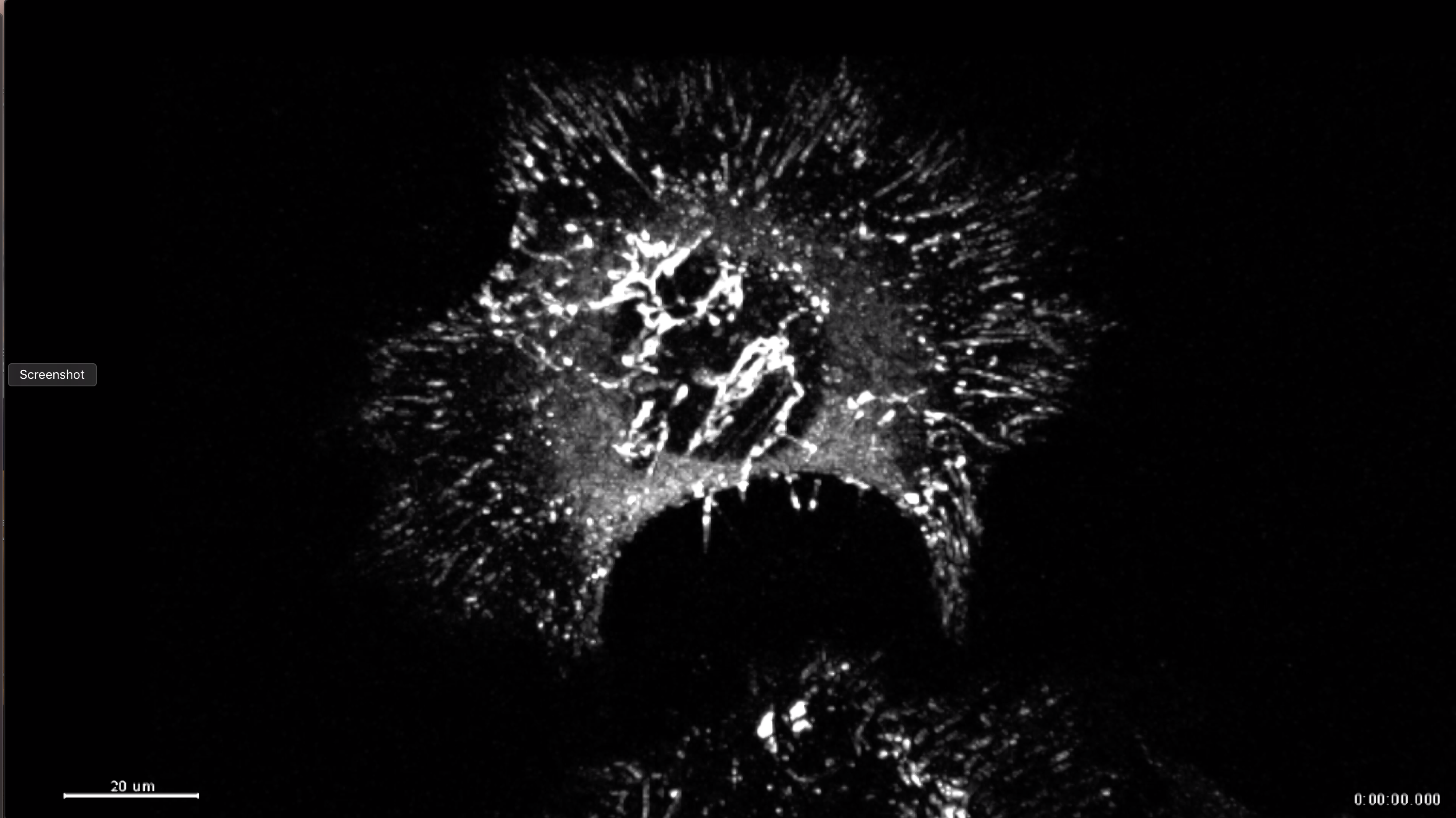
Fibronectin matrix formation in mouse embryo fibroblasts. Arrows point to fibril initiation sites. Photo Credit: Darshika Tomer
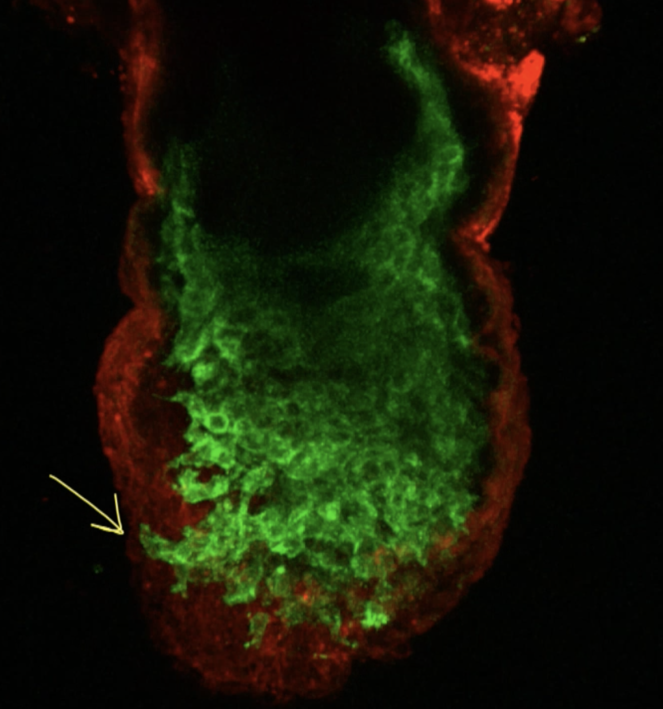
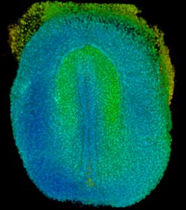
Migration of Mesp1-lineage mesoderm in E7.5 mouse embryo. Arrows point to migrating mesodermal cells. Photo Credit: Cecilia Arriagada