
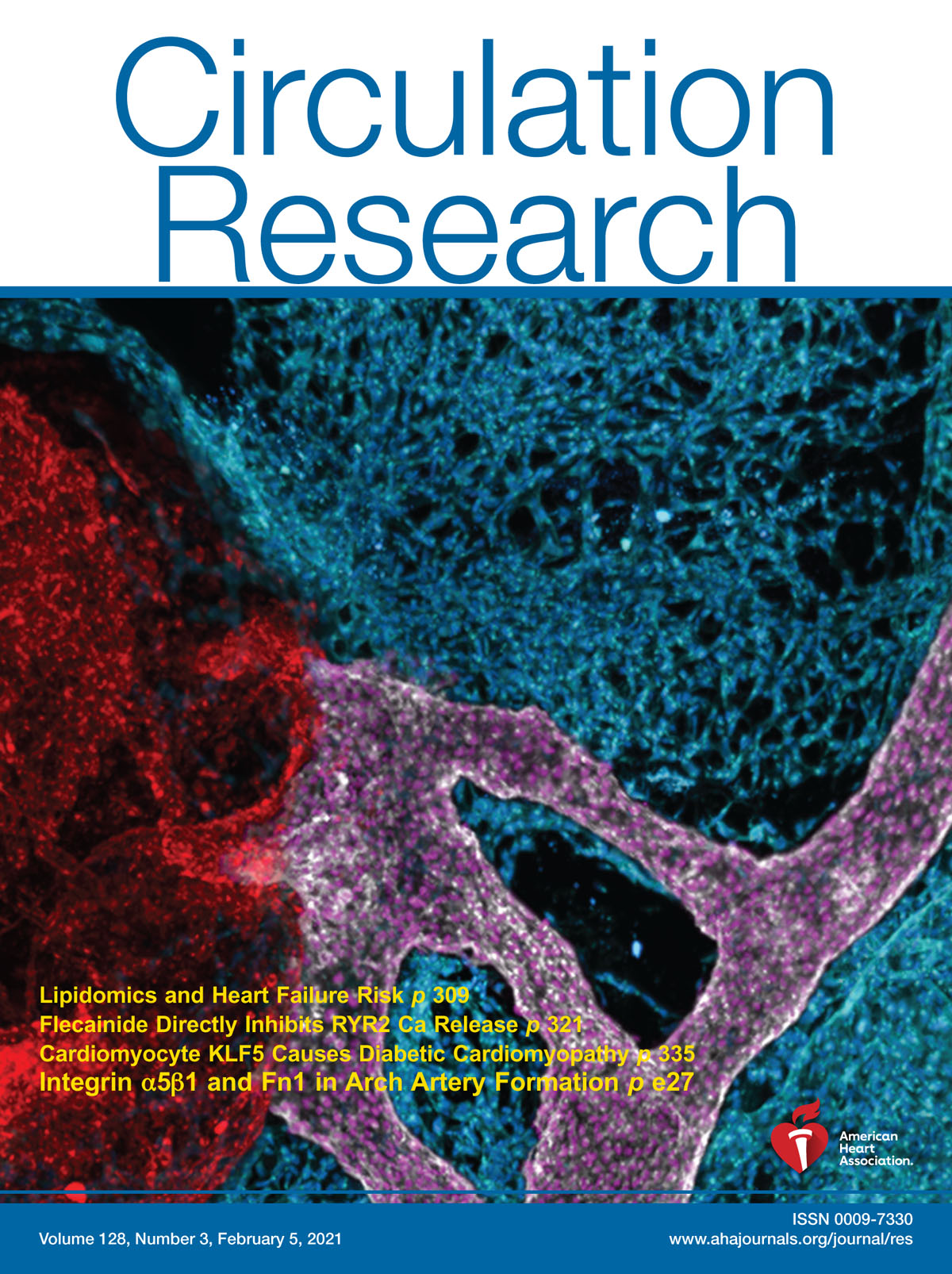
Buffering Mechanism in Aortic Arch Artery Formation and Congenital Heart Disease. Circulation Research Volume 134Issue 1010 May 2024Pages e112-e132, DOI: https://doi.org/10.1161/CIRCRESAHA.123.322767

This cover image was generated by AnnJosette Ramirez, Christina Vyzas, and Sophie Astrof:

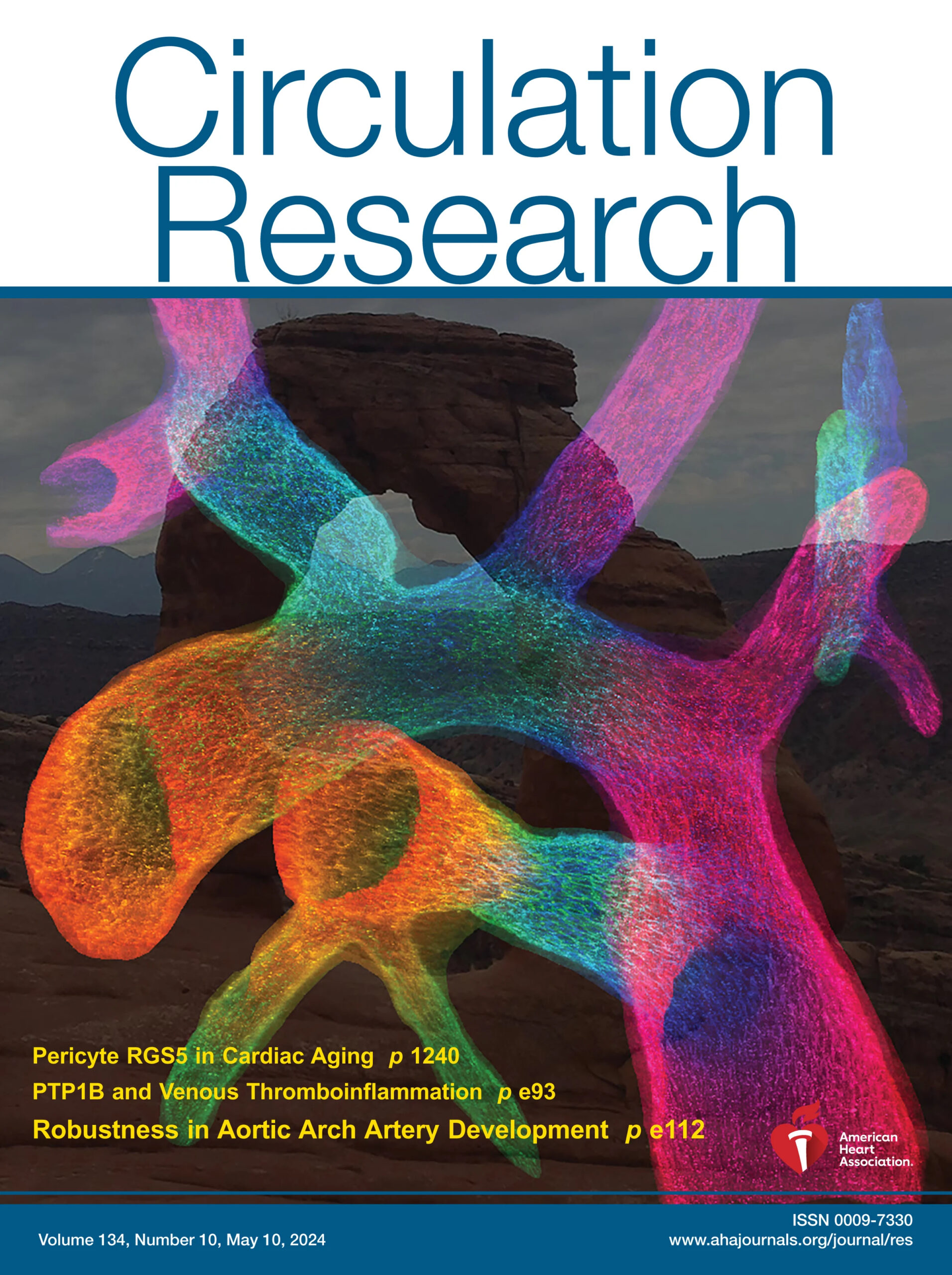
A 900-micron-thick vibratome section from a 14.5-day-old mouse embryo, containing the mature embryonic aortic arch artery tree with its branches, the ductus arteriosus, and their connecting vessels, was labeled using an antibody to Pecam-1 and imaged by confocal microscopy. The three-dimensional reconstruction of confocal slices was depth-color-coded and overlayed onto an image of the Delicate Arch, an iconic geological formation in Arches National Park, Utah, USA. The embryonic delicate arch, the 4th pharyngeal arch artery, and its derivative, the aortic arch, are the focus of the paper by Ramirez et al.

Meet the first author

Listen to the podcast @DiscoverCircRes

Cell-Extracellular Matrix Interactions Play Multiple Essential Roles in Aortic Arch Development

Pharyngeal arch artery formation is a multistep process entailing the dynamic contribution of SHF-derived ECs to pharyngeal arches, the remodeling of endothelial plexus into the PAAs, and the remodeling of the PAAs into the aortic arch artery and its major branches. Cell-ECM interactions regulated by integrin α5β1 and Fn1 play essential roles at each of these developmental stages.

See article by Warkala et al DOI: 10.1161/CIRCRESAHA.120.318200

See editorial by Robert Kelly DOI: 10.1161/CIRCRESAHA.121.318669

Meet the first author

Shape and position of the node and notochord along the bilateral plane of symmetry are regulated by cell-extracellular matrix interactions

The node and the notochord are positioned along the midline – the plane of bilateral symmetry in developing vertebrate embryos. The stereotypical shape and placement of these structures are essential for embryonic development. We show that cell-extracellular matrix interactions regulate the shape and orientation of the node along the plane of bilateral symmetry in the mouse and that this in turn regulates the placement of the notochord as well as its slender, cohesive shape. See the article by Pulina et al. (DOI: 10.1242/bio.20148243).

Essential roles of fibronectin in the development of the left-right embryonic body plan

We discovered that FN is required for the establishment of the asymmetric gene expression pattern in early mouse embryos by regulating morphogenesis of the node, while cellular fates of the nodal cells, canonical Wnt, and Shh signaling within the node were not perturbed by the absence of FN. Our studies demonstrate the requisite role for a structural ECM protein and its integrin receptor in the development of the left-right axis of asymmetry in vertebrates. See the article by Pulina et al. DOI: 10.1242/bio.20148243

Direct Test of Potential Roles of EIIIA and EIIIB Alternatively Spliced Segments of Fibronectin in Physiological and Tumor Angiogenesis

Upregulation of fibronectin and smooth muscle actin around tumor blood vessels. MOLECULAR AND CELLULAR BIOLOGY, Oct. 2004, p. 8662–8670 Cover photograph (Copyright © 2004, American Society for Microbiology. All Rights Reserved.): See article by Astrof et. al., DOI: 10.1128/MCB.24.19.8662-8670.2004